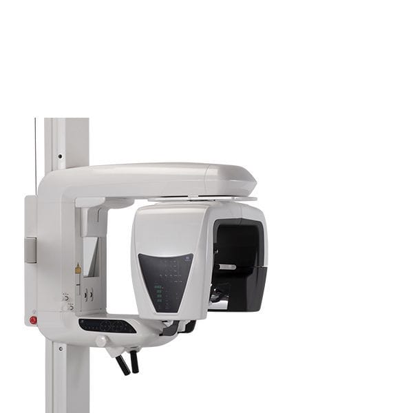
Veraview 3D R100
Veraview 3D R100
SKU:3DR100
Couldn't load pickup availability
The Veraviewepocs 3D R100 offers unique MORITA image quality for every dental practice. This unit has revolutionized 3D imaging and continues to set standards. Superior image quality in 3D and 2D, the MORITA-exclusive Panoramic Scout function, and the MORITA-exclusive Reuleaux image format, are just a few examples. In addition, there are features such as eight selectable exposure areas, an automatic exposure for panorama shots, and innovative techniques for automatic dose reduction.
Exclusive: Panoramic Scout for images of limited areas
With its MORITA-exclusive Panoramic Scout function, the Veraviewepocs allows you to easily and reliably define and set the right target area for small field CBCTs.
As a basis for this, you use an existing panoramic image. On the computer, just select the region in question and mark it with a small square. At the touch of a button, the device automatically moves to the correct position and is ready to acquire the CBCT image. The benefit for you: you create the image stress-free and right away in the region you are interested in. The benefit for your patient: A lower dose due to the limited area and also less stress when taking the image.
Two-directional Scout
After initial positioning is accomplished by the 3 positioning laser beams, 2-directional X-ray images can be taken to confirm that the position is accurate. If it is not, simply adjust the position of the image on the computer by placing the cursor at the center of the region of interest.
Direct Positioning with Laser Beams
Positioning laser beams set the patient’s position and align the region of interest manually. First, the patient’s initial position is set using the 3 laser beams. Then, 2 additional laser beams are aligned to the region of interest. The C-arm will automatically move to the right position.
Six Fields of View:
Veraviewepocs 3D R100 offers a total of 6 exposure areas from 40 x 40 mm up to 100 x 80 mm for various diagnostic needs. The full arch scan captures the maxilla and/or the mandible with the equivalent of 100 mm in diameter and two height options of 50 or 80 mm. Its full arch capability, reduced dose, and exceptional clarity are ideal features for implant planning and oral surgery.
Ø 100 x H 80 mm (3D Reuleaux Full Arch FOV)
Ø 100 x H 50 mm (3D Reuleaux Full Arch FOV)
Ø 80 x H 80 mm
Ø 80 x H 50 mm
Ø 40 x H 80 mm
Ø 40 x H 40 mm
Within the 100 mm diameter exposure range MORITA’s unique 3D system replaces the typical cylindrical shape with a convex triangle shape - the Reuleaux. The generated unique FOV encompasses the entire dental arch (with upper jaw and lower jaw) and simultaneously excludes all other areas of image acquisition, that are out of interest. The benefit of this MORITA-exclusive function: Imaging of the entire dental arch can be achieved with less X-ray dose.
Blue line indicates full arch FOV, equivalent to ∅ 100 mm
Innovative 3D Reuleaux Full Arch FOV reduces dose.


